
Tracheal stenosis is a condition characterized by a narrowing of the trachea, which can lead to severe breathing difficulty, coughing, and recurrent respiratory infections. It’s often a complication arising from prolonged intubation, tracheostomy, or can be due to inflammatory diseases such as tuberculosis and trauma, or tumors. Patients with tracheal stenosis typically report a progressive worsening of breathlessness, especially during exertion, and a high-pitched wheezing (stridor), signalling the need for medical evaluation.
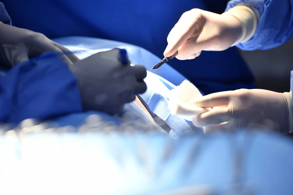
Diagnosis involves bronchoscopic examination of the airways, pulmonary function tests, and imaging studies like CT scans to determine the stenosis’ severity and location. This stenosis can be “simple” or “complex”. “Simple stenoses” can be managed with bronchoscopic techniques (balloon tracheoplasty). or stent placement), however, “Complex stenosis” requires surgical intervention for long-term resolution, if bronchoscopic treatment (balloon tracheoplasty or stent placement) fails. Surgery typically involves removal the narrowed section of the trachea and reconstruct the airway (surgical resection and anastomosis).
Dr. Mohan Venkatesh Pulle, as an experienced tracheal (airway) surgeon, offers specialized treatment for tracheal stenosis, employing advanced techniques to optimize patient recovery and ensure the restoration of normal breathing.
Tracheal Stenosis: Understanding a Narrowed Windpipe
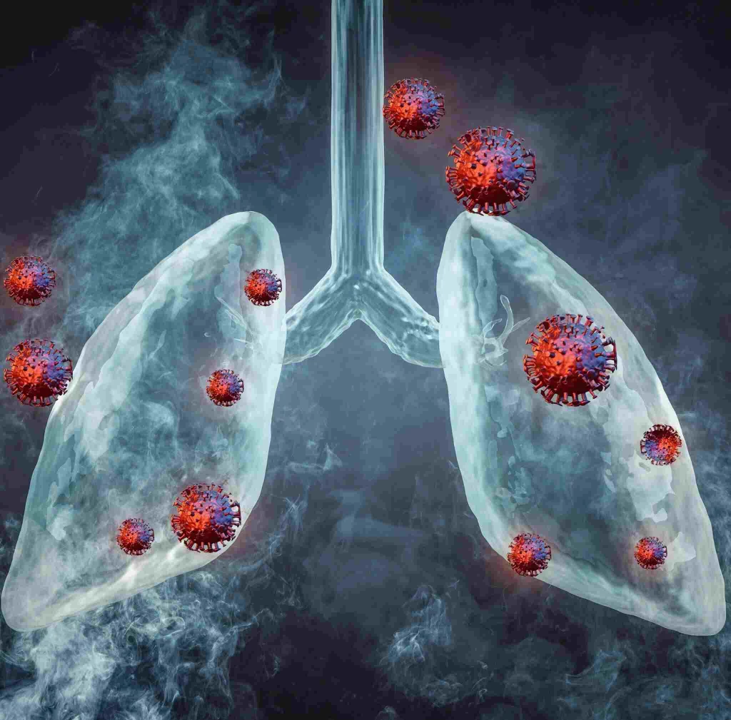
Tracheal stenosis is a medical condition characterized by the narrowing of the trachea, the main airway that connects the larynx (voice box) to the lungs. This narrowing can restrict the flow of air into and out of the lungs, leading to breathing difficulties and other respiratory symptoms. Tracheal stenosis can result from various causes, and understanding its pathophysiology, clinical presentation, diagnosis, and treatment is essential for effective management and improved patient outcomes.
What is Tracheal Stenosis?
Anatomy and Function of the Trachea
The trachea, also known as the windpipe, is a rigid, tube-like structure that starts just below the larynx and extends into the chest, where it divides into the right and left main bronchi, leading to the lungs. The trachea is composed of C-shaped cartilage rings connected by fibrous tissue and smooth muscle. This unique structure allows the trachea to remain open for airflow while still providing flexibility for swallowing and neck movement.
The primary function of the trachea is to transport air to and from the lungs during the process of breathing. It acts as a conduit for the exchange of oxygen and carbon dioxide, playing a vital role in the respiratory system.
Causes of Tracheal Stenosis
Tracheal stenosis can be classified as congenital or acquired, depending on its underlying cause:
- Congenital Tracheal Stenosis: This type of stenosis is present at birth and results from abnormal development of the trachea during fetal growth. It can be caused by factors such as genetic mutations, prenatal infections, or intrauterine trauma.
- Acquired Tracheal Stenosis: Acquired stenosis occurs after birth and can result from various factors, including:
a) Trauma: External injuries or internal trauma to the trachea, such as a direct blow to the neck or a penetrating injury, can cause scarring and narrowing of the airway.
b) Endotracheal Intubation: Prolonged intubation with a breathing tube, as is often done during surgery or in the intensive care unit, can lead to tracheal damage and subsequent stenosis.
c) Infections and Inflammatory Conditions: Conditions such as prolonged intubation with a breathing tube, as is often done during surgery or in the intensive care unit, can lead to tracheal damage and subsequent stenosis.
d) Autoimmune Disorders: Certain autoimmune diseases, such as relapsing polychondritis, can cause inflammation of the tracheal cartilage, leading to stenosis.
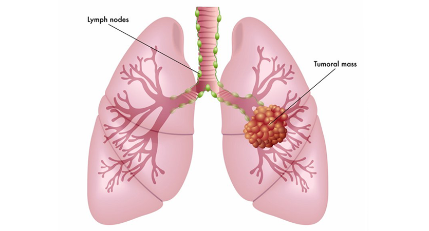
e) Tumors: Benign or malignant growths in or around the trachea can compress the airway and cause narrowing.
f) External Compression:** Compression of the trachea by structures outside the airway, such as an enlarged thyroid gland or a mediastinal mass, can also result in stenosis.
Clinical Presentation
The symptoms of tracheal stenosis can vary depending on the degree of narrowing and the underlying cause. Common signs and symptoms may include:
- Shortness of breath (Dyspnea): Breathing becomes difficult and may be accompanied by a feeling of suffocation.
- Wheezing: A high-pitched whistling sound during breathing due to narrowed air passages.
- Stridor: A harsh, noisy breathing sound that occurs during inspiration and is often a sign of significant tracheal narrowing.
- Chronic Cough: A persistent cough may be present, especially if there is irritation of the airway.
- Recurrent Respiratory Infections: Tracheal stenosis can compromise the body’s ability to clear mucus and debris, leading to an increased risk of respiratory infections.
- Cyanosis: Bluish discoloration of the skin and mucous membranes due to reduced oxygenation of the blood.
- Exercise Intolerance: The limited airflow can lead to fatigue and shortness of breath with physical activity.
In severe cases, tracheal stenosis can lead to life-threatening respiratory distress, especially during periods of exertion or stress.
Diagnosis and Evaluation
The diagnosis of tracheal stenosis starts with a thorough medical history and physical examination, paying close attention to respiratory symptoms and risk factors. Diagnostic tests and imaging studies may be ordered to confirm the diagnosis and assess the severity of the stenosis:
- Pulmonary Function Tests (PFTs): These tests measure lung function and airflow, helping to identify any significant respiratory abnormalities.
- Chest X-ray: X-ray imaging can provide initial information about the tracheal anatomy and any potential masses or compressions.
- Computed Tomography (CT) Scan: CT scans offer detailed cross-sectional images of the trachea, allowing the physician to assess the degree and extent of stenosis.
- Bronchoscopy: This is a crucial diagnostic tool for directly visualizing the trachea and airways. A flexible bronchoscope is inserted through the nose or mouth and passed into the trachea, allowing the physician to visualize any stenotic areas and take tissue samples if needed.
Classification of Tracheal Stenosis
Tracheal stenosis is classified based on its location and extent within the trachea:
- Subglottic Stenosis: Narrowing of the trachea just below the level of the vocal cords (glottis). This type can result from intubation injury or autoimmune conditions.
- Tracheal Stenosis: Stenosis involving any part of the trachea, from the level of the glottis to the division of the main bronchi.
- Tracheobronchial Stenosis: Narrowing that extends into the bronchi, the smaller airways leading to the lungs.
Treatment Options
The management of tracheal stenosis depends on its severity, location, and underlying cause. Treatment options include:
- Medical Management: Mild cases of tracheal stenosis may be managed conservatively with medications to alleviate symptoms and treat any underlying infections or inflammation.
- Endoscopic Interventions: For certain types of tracheal stenosis, endoscopic techniques may be used to dilate the narrowed airway and place stents to keep it open.
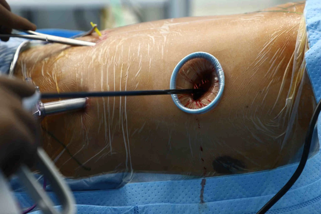
- Surgical Interventions: In more severe cases or when endoscopic methods are not sufficient, surgical procedures may be necessary to repair or reconstruct the trachea. Surgical options include tracheal resection and anastomosis, laryngotracheal reconstruction, or tracheal transplantation.
- Tracheostomy: In cases where the trachea is severely compromised and other interventions are not feasible, a tracheostomy may be performed. This involves creating a surgical opening in the neck to insert a breathing tube directly into the trachea.
Prognosis and Complications
The prognosis for individuals with tracheal stenosis depends on the extent of the narrowing, the presence of any associated medical conditions, and the response to treatment. Mild cases may be effectively managed with conservative measures, while more severe stenosis may require surgical interventions.
Complications of tracheal stenosis can include recurrent respiratory infections, respiratory distress, and worsening respiratory function. Prompt diagnosis and appropriate management are crucial for preventing complications and improving long-term outcomes.
Rehabilitation and Follow-up
After treatment for tracheal stenosis, rehabilitation and follow-up care are essential for optimizing recovery and monitoring the airway’s health. Individuals may require pulmonary rehabilitation to improve lung function and enhance exercise tolerance. Regular follow-up visits with healthcare providers are necessary to assess the airway’s patency and address any potential complications or recurrence.
Emerging Therapies and Research
Advancements in medical technology and research continue to drive the development of new therapies for tracheal stenosis. Innovative approaches, such as tissue engineering and regenerative medicine, hold promise for enhancing tracheal reconstruction and improving outcomes for individuals with severe stenosis.
Prevention and Risk Reduction
While congenital tracheal stenosis may not be preventable, certain measures can reduce the risk of acquired stenosis:
- Proper Intubation Techniques: Healthcare providers should adhere to best practices for endotracheal intubation and closely monitor patients undergoing prolonged intubation to minimize the risk of tracheal damage.
- Early Detection and Treatment: Prompt identification and management of respiratory infections, autoimmune conditions, and other potential risk factors can help prevent the development of tracheal stenosis.
Frequently asked questions
Doctor, what exactly is Tracheal Stenosis?
See, Tracheal Stenosis is basically narrowing of your windpipe. Think of it like water pipe becoming narrow at some point. This makes breathing difficult because less air can pass through.
What caused this condition in my case?
Most common causes are prolonged intubation, previous throat infection, or injury. Sometimes it happens after tracheostomy also. In your case, [specific cause] seems to be main reason.
Is this life-threatening condition?
It can be serious if left untreated, but nowadays we have very good treatment options. Key is early diagnosis and proper treatment. Don’t worry too much – we’ll handle this properly.
What treatment options are available?
We have several options – endoscopic dilation, laser treatment, or surgery depending on severity. In your case, I’m thinking [specific treatment] might work best. We’ll discuss pros and cons in detail.
Will I need surgery?
Not everyone needs surgery. We first evaluate severity and length of narrowing. Sometimes simpler procedures work well. But if surgery is needed, we use advanced minimally invasive techniques.
How long will recovery take?
Recovery timeline varies. After endoscopic procedure, you might go home next day. For surgery, usually 5-7 days in hospital. Full recovery takes 4-6 weeks. But breathing improvement you’ll notice quite quickly.
Will my voice be affected?
There might be temporary voice changes, but usually returns to normal. We take special care to protect voice box during treatment. If needed, we’ll arrange speech therapy.
Can this problem come back after treatment?
There’s small chance of recurrence, but we’ll teach you exercises and give medications to prevent this. Regular follow-up helps catch any problems early.
What precautions should I take after treatment?
Main things – no smoking absolutely, avoid dusty environments, do breathing exercises regularly, complete all medicines. Also, very important to come for follow-up appointments.
When can I return to normal activities?
Not everyone needs surgery. We first evaluate severity and length of narrowing. Sometimes simpler procedures work well. But if surgery is needed, we use advanced minimally invasive techniques.
What about exercise and sports?
Start with gentle walking first week after discharge. Then gradually increase as comfortable. No heavy exercise for 6 weeks. Swimming is excellent once wound heals completely.
How will I know if treatment is working?
You’ll notice breathing becomes easier, less noisy. Exercise capacity will improve. We’ll do regular breathing tests to measure improvement objectively.
What symptoms should worry me after treatment?
Watch for increased breathing difficulty, noisy breathing, fever, or throat pain. If any of these occur, contact us immediately. But don’t panic – complications are rare.
Will I need long-term medications?
Initially yes, but usually temporary. Main medicines are for preventing inflammation and infection. Some patients might need inhalers for few months.
How can I prevent this from happening again
Follow-up regularly, complete all treatments, do breathing exercises, maintain good throat hygiene. If you get throat infections, treat them properly and timely.
Important Note: “Remember, every patient’s condition is bit different. We’ll customize treatment plan for you. My team and I are always available if you have concerns. Good communication helps in better recovery.”
What causes this narrowing?
Most common causes are prolonged intubation, previous throat surgery, wind-pipe tumors or some injury to throat. Sometimes it can happen from infections also. In your case, we’ll need to find exact cause.
How serious is this condition?
It can be quite serious if left untreated because it affects your breathing. But don’t worry too much – we have very good treatment options nowadays. Success rates are quite encouraging.
What are my treatment options?
Depending on severity, we have different options – sometimes we can do endoscopic dilation, sometimes we need surgery. Please contact me and send your reports, we will decide the appropriate plan for you.
Will I need surgery immediately?
Not always immediate. First we’ll do proper evaluation with breathing tests and scans. Sometimes we try conservative treatment first. We’ll decide best approach for your condition.
How long will surgery take?
Usually takes 2-3 hours. In surgery, the diseased portion of the trachea is removed and the normal tracheal is re-connected to the normal trachea part. In simple words, if a portion of the water-pipe is damaged you are removing that portion and re-joining the normal portions. As I said, this looks very simple but requires a very good experienced team to perform it very diligently. We use advanced techniques to repair stenosis and we have the highest experience in managing such problems.
What about the recovery period?
Typically patients stay in hospital for 5-7 days. Complete recovery takes about 4-6 weeks. But you’ll start noticing breathing improvement immediately after surgery.
What complications should I watch for?
Main things to watch – breathing difficulty, excessive coughing, fever, or throat pain. If you notice any of these, contact us immediately. But complications are quite rare with proper care.
When can I return to work?
Most patients return to desk job in 3-4 weeks. Physical work might need 6-8 weeks. We’ll assess your recovery and guide accordingly.
Can I exercise after treatment?
Can start with gentle walking from next day after surgery. Gradually increase activity. No heavy exercise for 2-3 months. We’ll give you a proper exercise plan based on your recovery.
What about diet restrictions?
For few days after surgery all food will be given through a pipe,not from mouth. This is because the swallowing mechanism will take some time to come to normal state after surgery. Till the time it recovers, this tube feeding will help you to maintain the nutrition. Within a week or 10 days, once the normal swallowing mechanism recovers, we can start oral diet. Initially soft diet is better. Avoid very hot or cold foods. Small, frequent meals are good. We’ll give detailed diet instructions before discharge.
How often will I need follow-up?
First month weekly, then monthly for 3 months. After that, every 6 months if everything stable. But if any problems develop, contact immediately.
Can this problem come back?
Recurrence is a rarity after proper surgery if all the surgical principles are followed. Regular follow-up helps catch any problems early. Following post-treatment instructions is very important.
What lifestyle changes are needed?
No smoking absolutely, avoid dusty environments, maintain good throat hygiene. Regular breathing exercises help. We’ll teach you specific techniques.